


| ISM real art exhibition – 2021 |
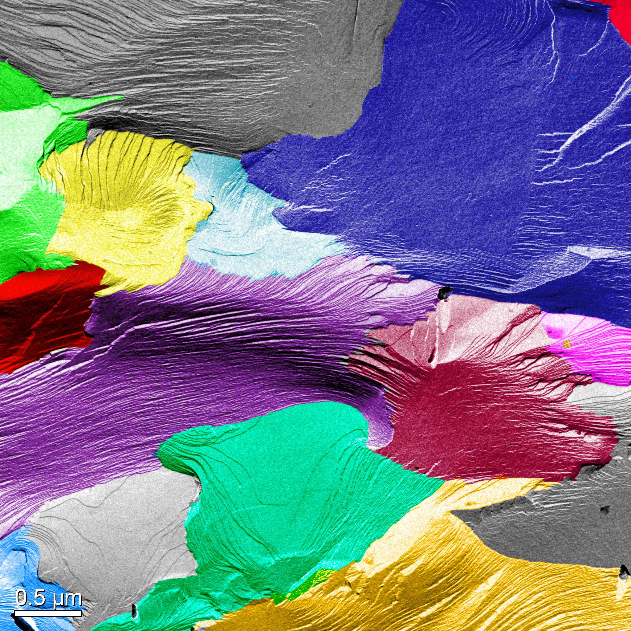 | Fight of colors Ellina Kesselman / Technion TEM Micrograph of a Freeze-fracture replica, Tecnai T-12 TEM, artificial coloring. PI: Prof. Ishi Talmon, Technion. |
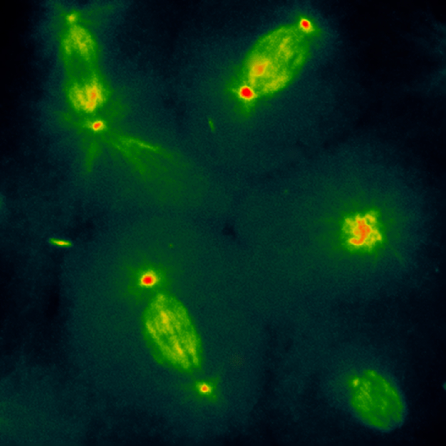
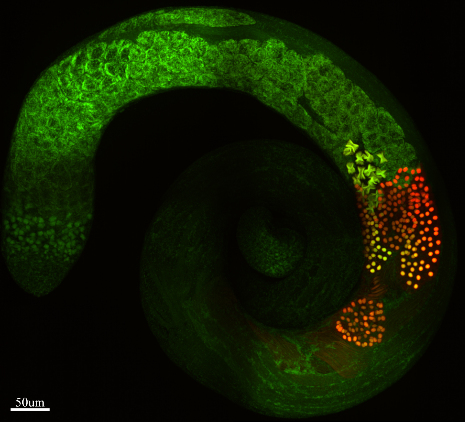 | Testi-color Alina Kolpakova / Weizmann Institute Drosophila testis. Expression pattern of the somatic (Marf, green) and the testis-specific (Fzo, red) Mitofusins, visualizing mitochondrial morphological changes during spermatogenesis. The image was obtained using Dragonfly spinning disc confocal microscope. |
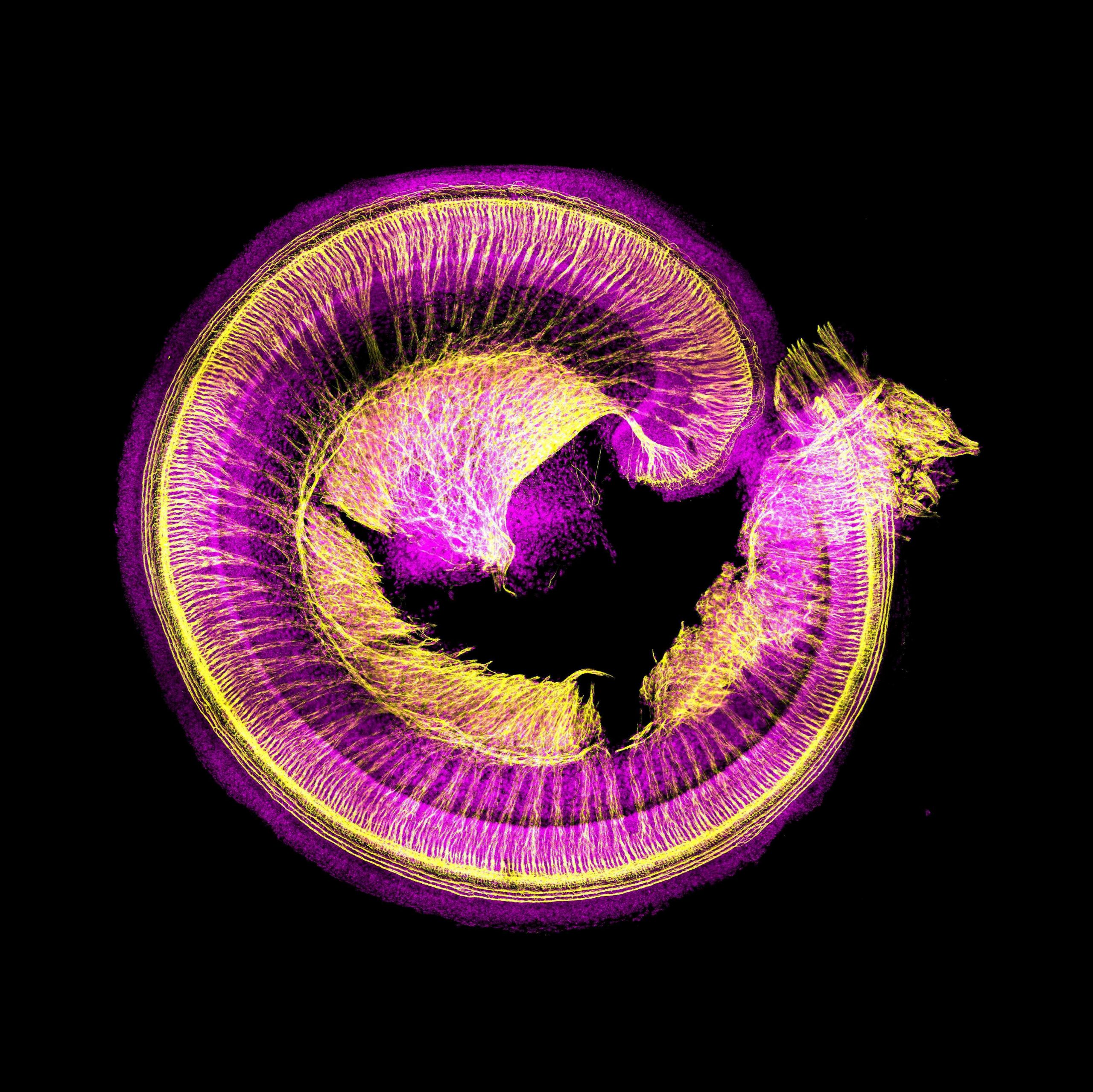 | Spiral ganglion neurons Shahar Taiber / Tel-Aviv University Confocal image of a mouse neonatal organ of Corti. Spiral ganglion neurons are stained in yellow (beta3-tubulin) and nuclei are stained in pink (DAPI) |
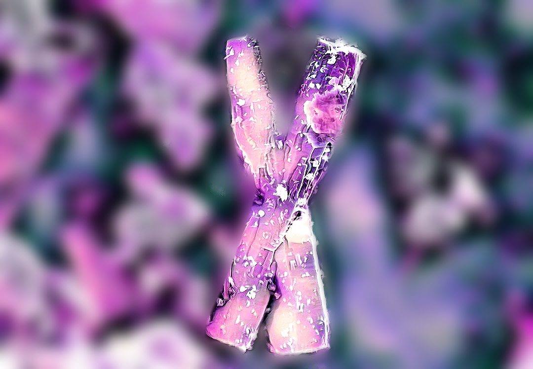 | ZnO Chromosomes Shir Tabac / Technion Microscope micrograph of ZnO seed microrods observed at SEM that with an uncanny resemblance to a pair of chromosomes. Photo taken by EliyahuFarber. |
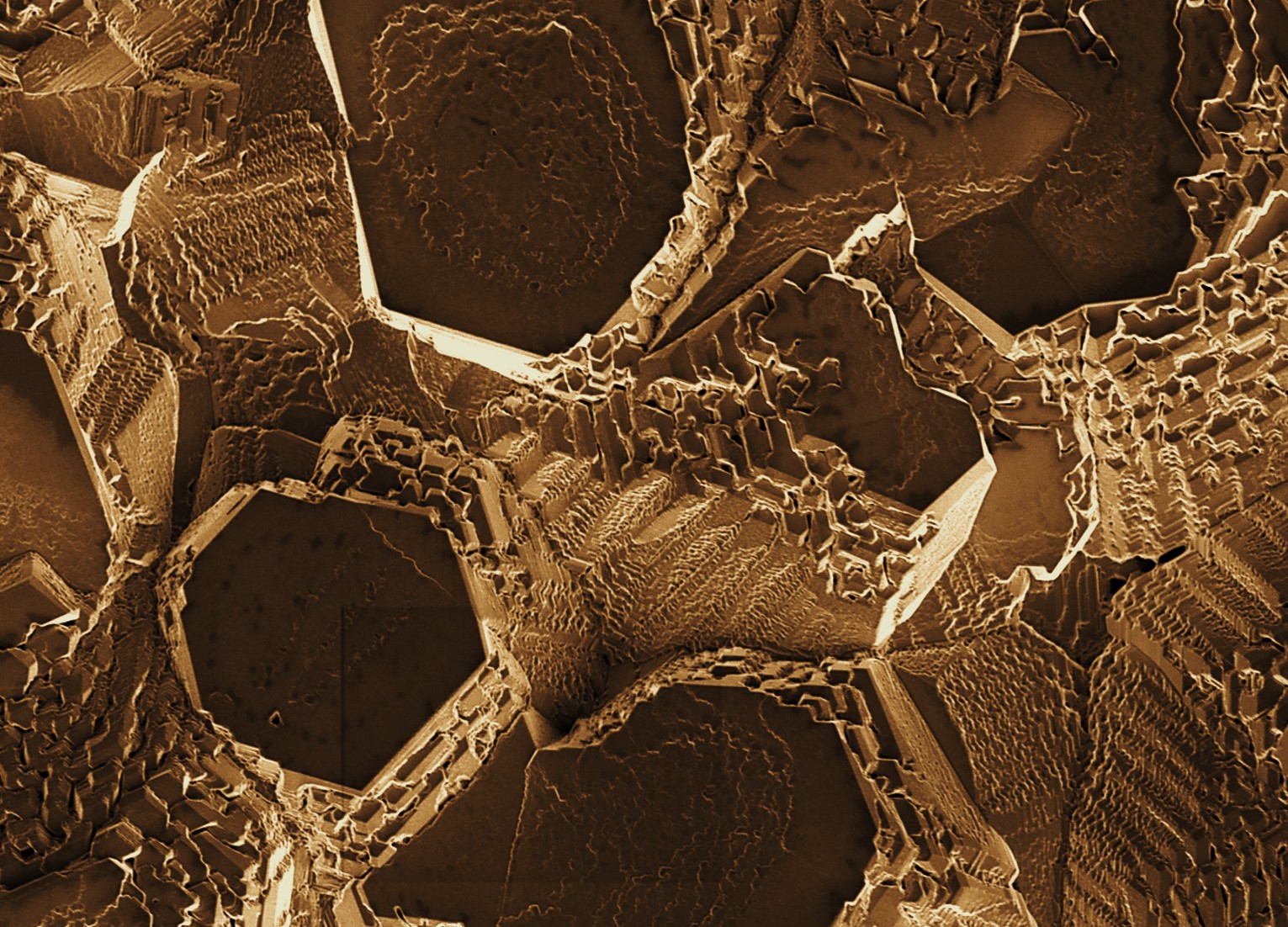
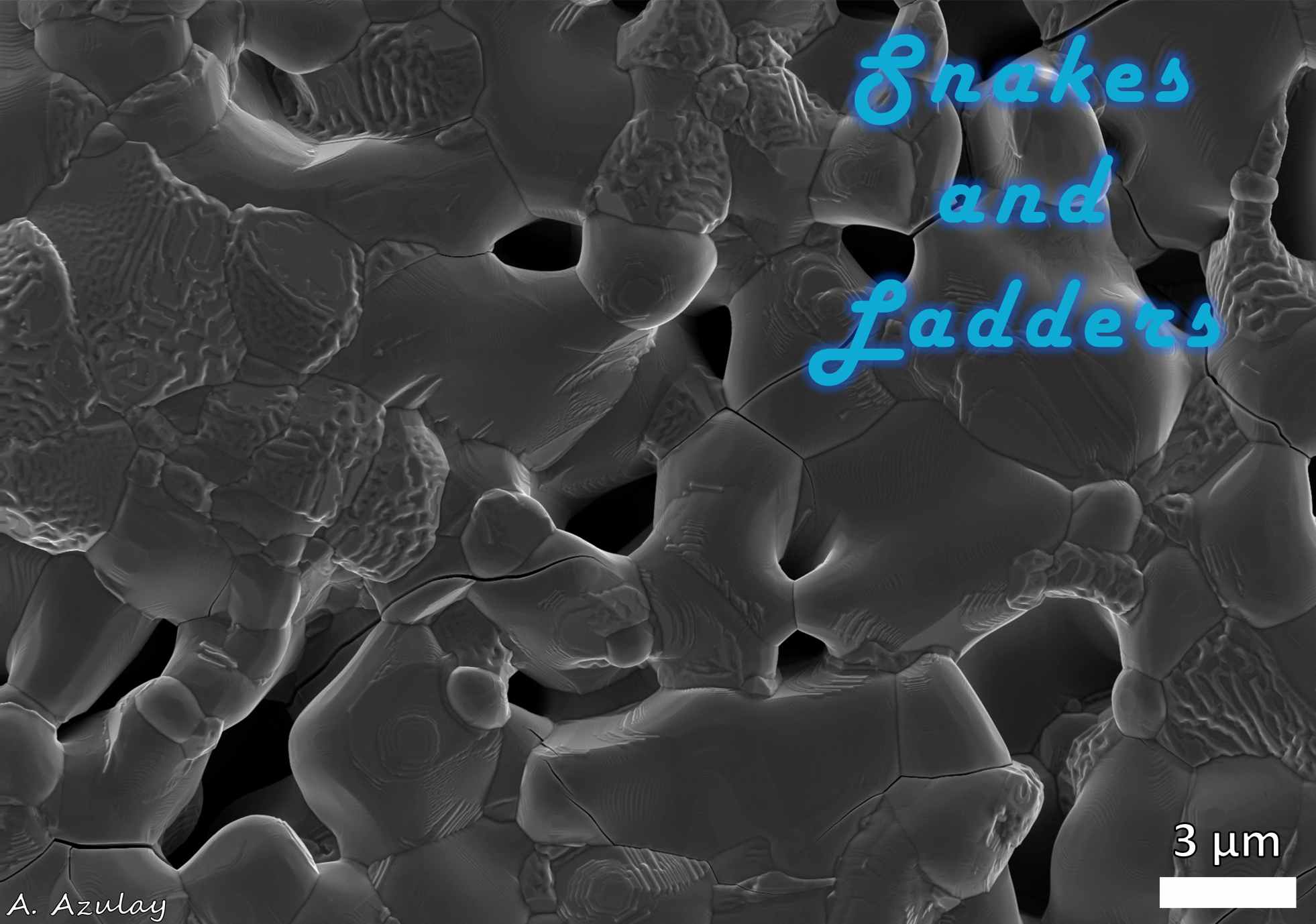 | Snakes-and-Ladders Amram Azulay / Technion Scanning electron microscopy SE micrograph taken from the surface of as-sintered polycrystalline Y-doped Ca2MnO4 sample for thermoelectric energy conversion. |
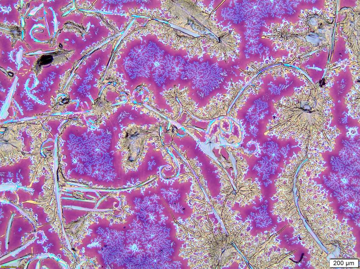
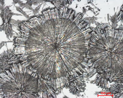
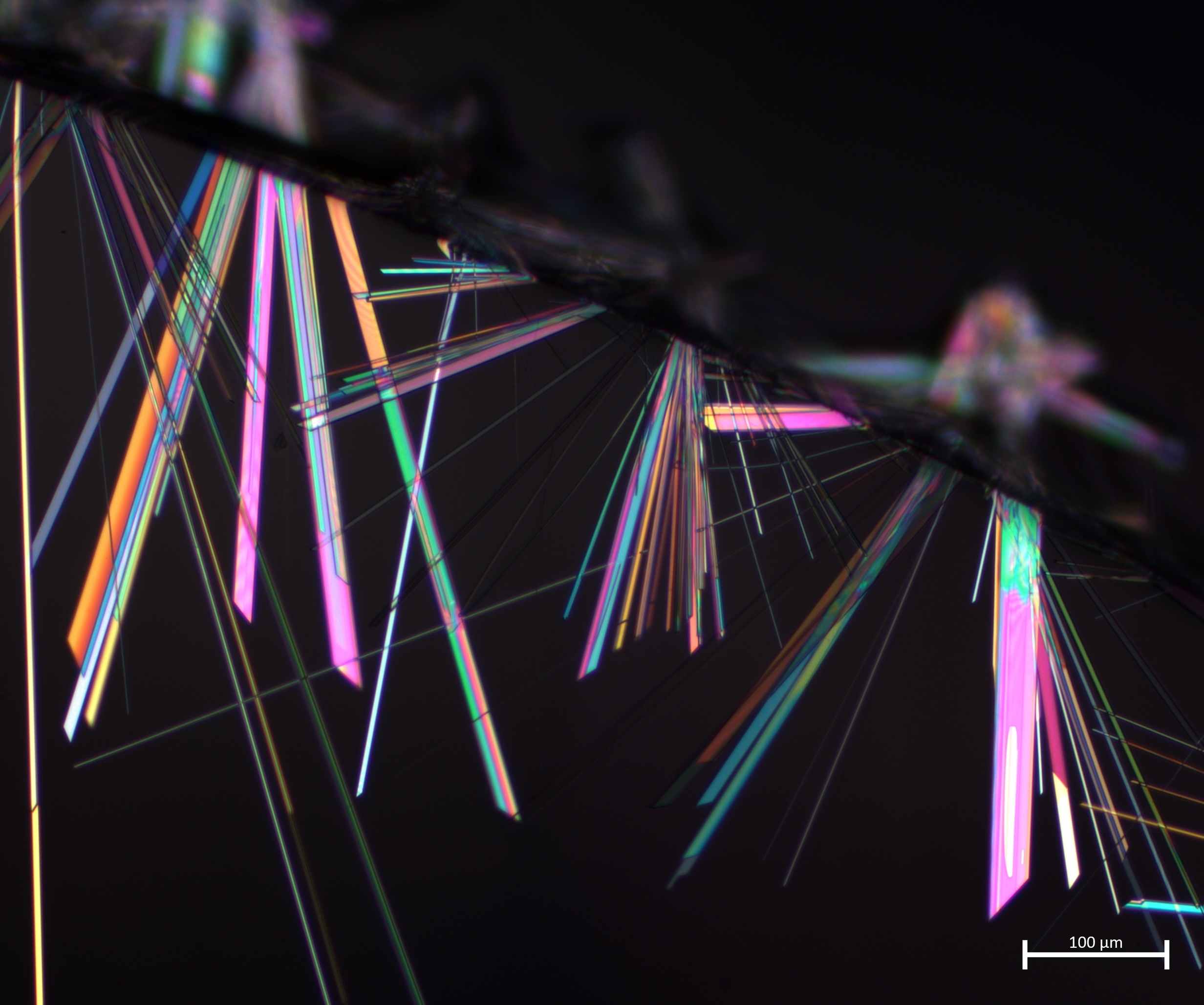 | Disco party Avital Wagner / Ben-Gurion University Polarizing microscope image of hypoxanthine monohydrate crystals. The colors in the image are created naturally by optical birefringence of the crystals. |
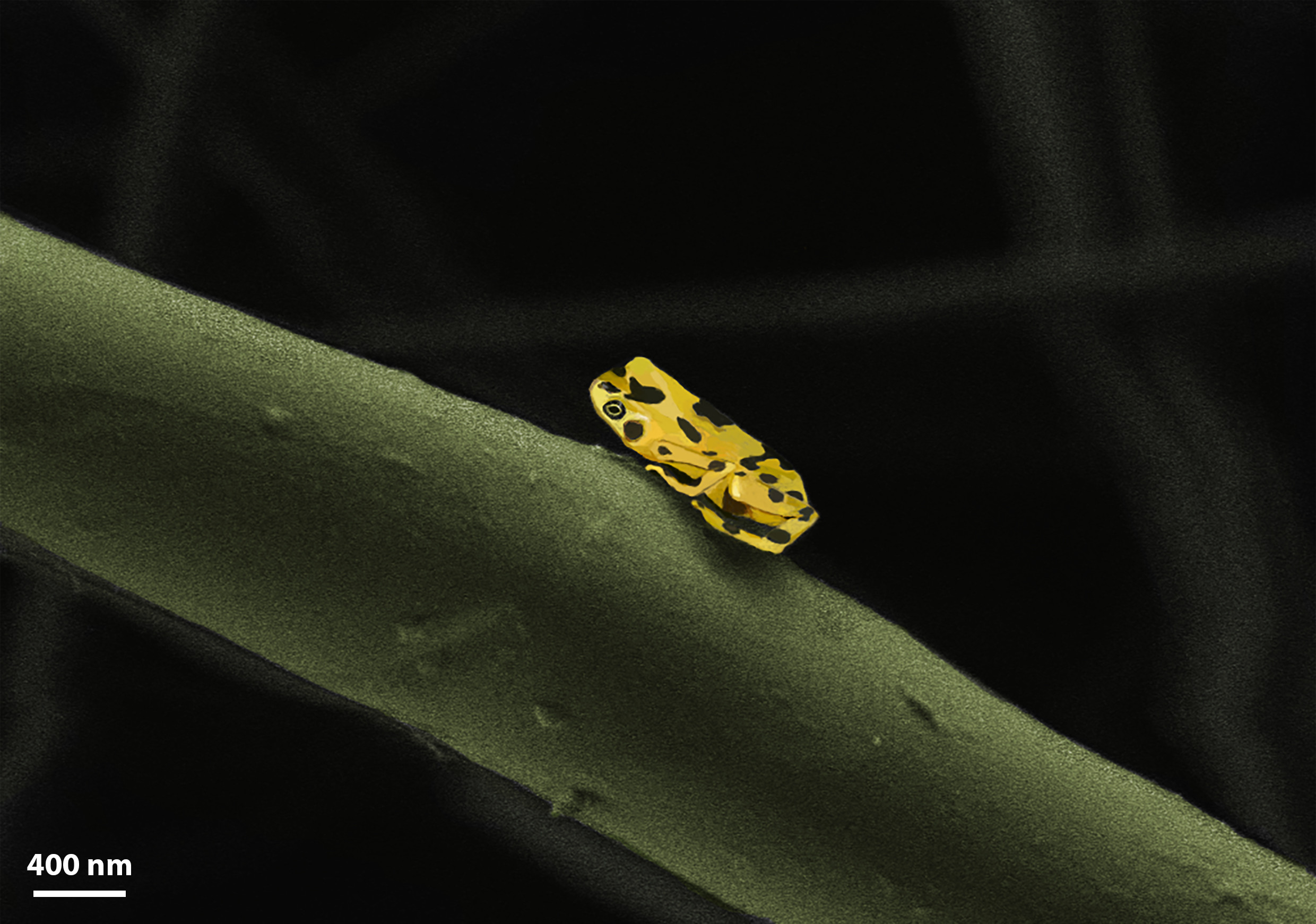 | Nano-frog Oren Elishav / Technion Painted SEM SE image of electrospun polyvinylpyrrolidone nanofiber contaminated with a nanoparticle in the shape of a frog. |
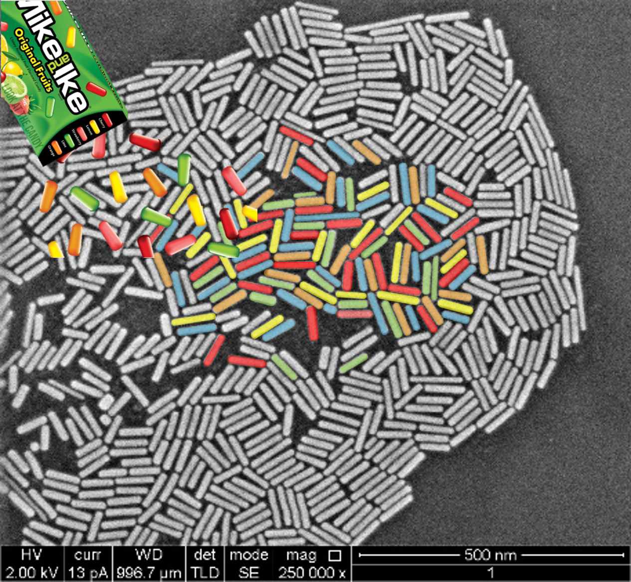 | "Mike & Like" limited edition Nethanel Friedman / Hebrew University Fabricated gold nanords demonstrated in TEM, mag: X250,0000. |
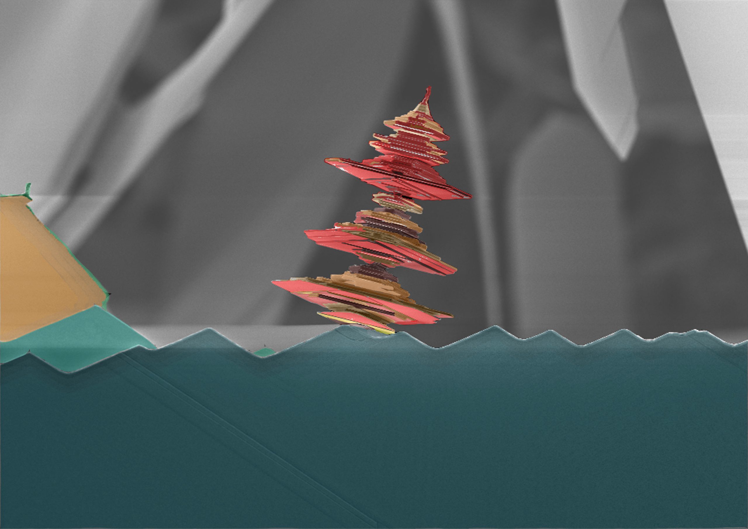 | Pagoda on Water Ella Sanders / Weizmann Institute Layers of microscopic lead iodide placed in different orientations by SEM surface imaging. |
 | Sweet Division Shai Adar Levor / Ben-Gurion University Image of dividing cells at zebrafish embryo in blastula stage. Imaged using spinning disk confocal microscope. Microtubules are shown. |
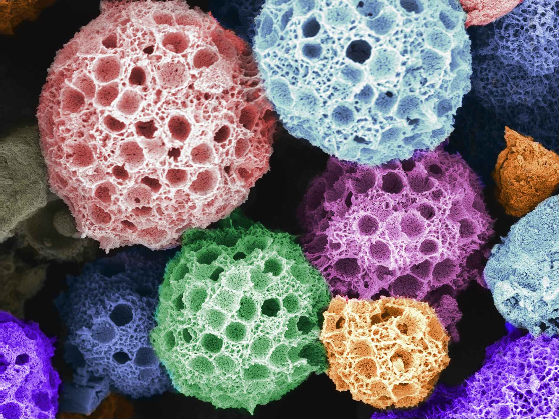 | Coral Yu Chen, Gal Radovsky & Ehud Gazit / Tel-Aviv University Scanning electron microscopy image with BSE detector showing carnosine-Zn supramolecular multi-porous microspheres. They were synthesized using the seeded emulsion assisted crosslinking method. The image shows the near-spherical of Carnosine-Zn, with an average diameter of ~5 μm, and uniformly distributed macroporous structures on the surface with a diameter of ~300 nm, as well as mesoporous structures formed between thescaffolds, with a mean size of 2-10 nm |
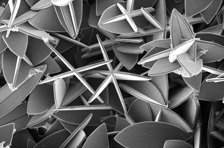
 | Spicules Neta Varsano / Weizmann Institute Artistic representation of ascidian spicules produced by Didemnidae a family of colonial tunicates. The SEM micrograph was taken by prof. Lowenstam Heinz – one of the pioneers in the field of biomineralization. |
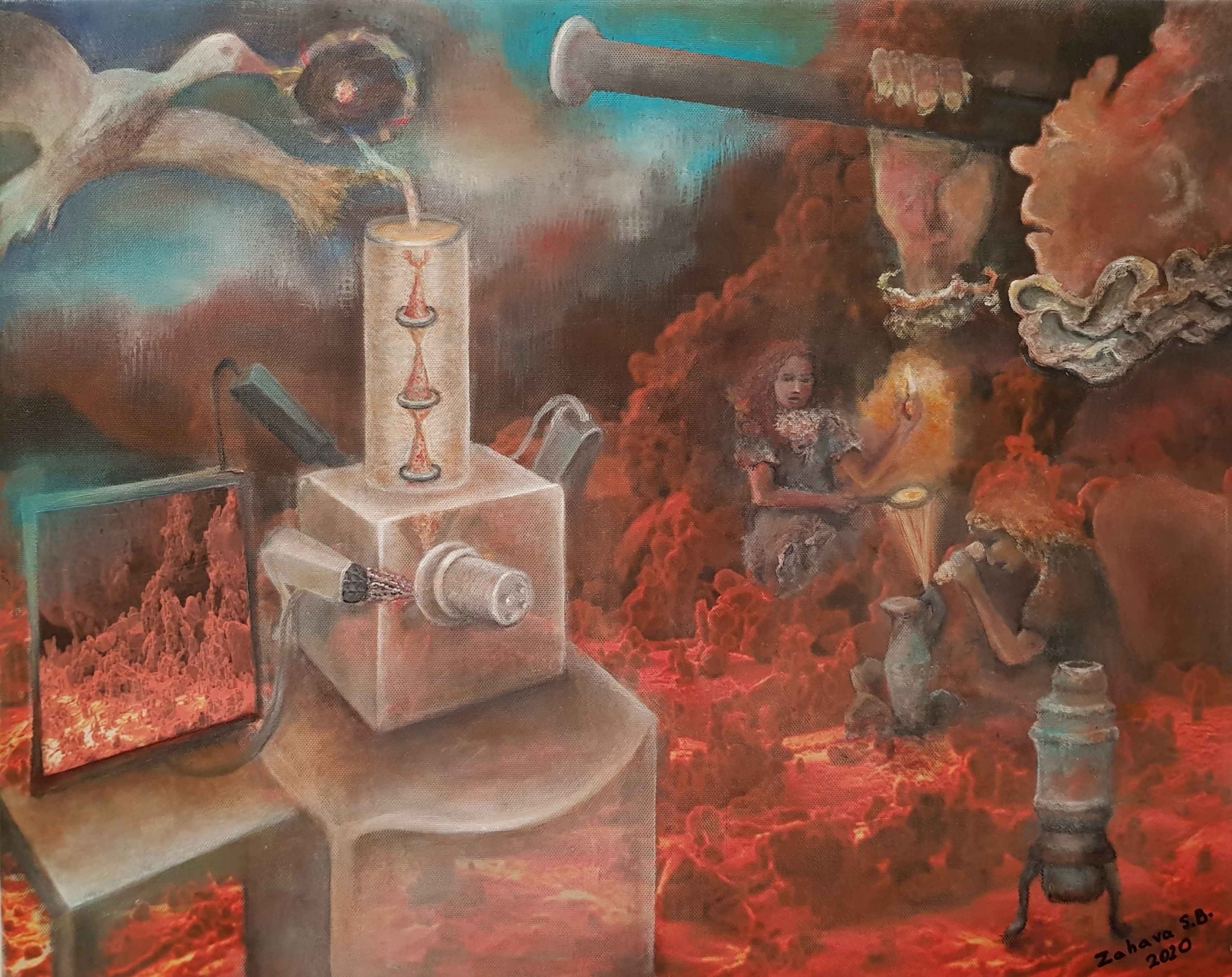 | History of Electron and Optical Microscopy Zahava Barkay / Tel-Aviv University Historical development of microscopy is shown in this acrylic on canvas painting: scanning electron microscopy including atomic model (on the left) and various optical tools including Galileo microscope (on the right). The image on the SEM screen provides the background for the whole scene. |
 | Winter Winds Rotem Zilberberg / Technion Spiral and fractal Si crystals surrounded by gold. Microscope – Olympus BX51. |
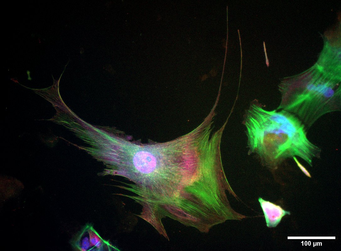 | Podocyte Bat Marina Safrigin / Bar-Ilan University Cell Bat. hiPSC derived podocyte stained by Hoechst for nuclei, phalloidin for actin, and synaptopodin immunofluorescence staining for actin-associated proteins. Optical Microscope X20 |
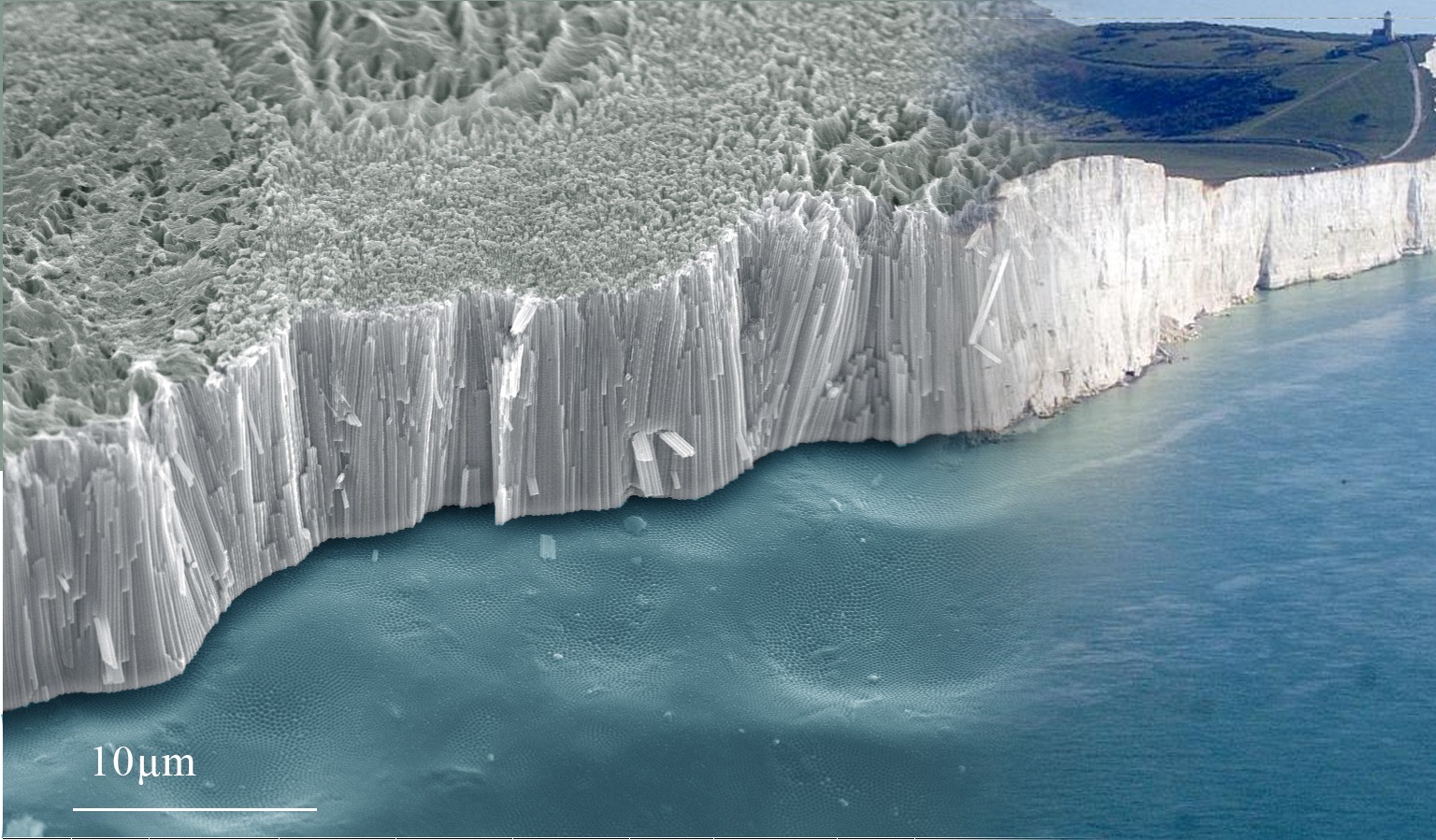 | White Cliffs of Dover Gil Bergman / Bar-Ilan University SEM Micrograph of TiO2 nanotubes cross section using a HR-SEM transforms into the White Cliffs of Dover. |
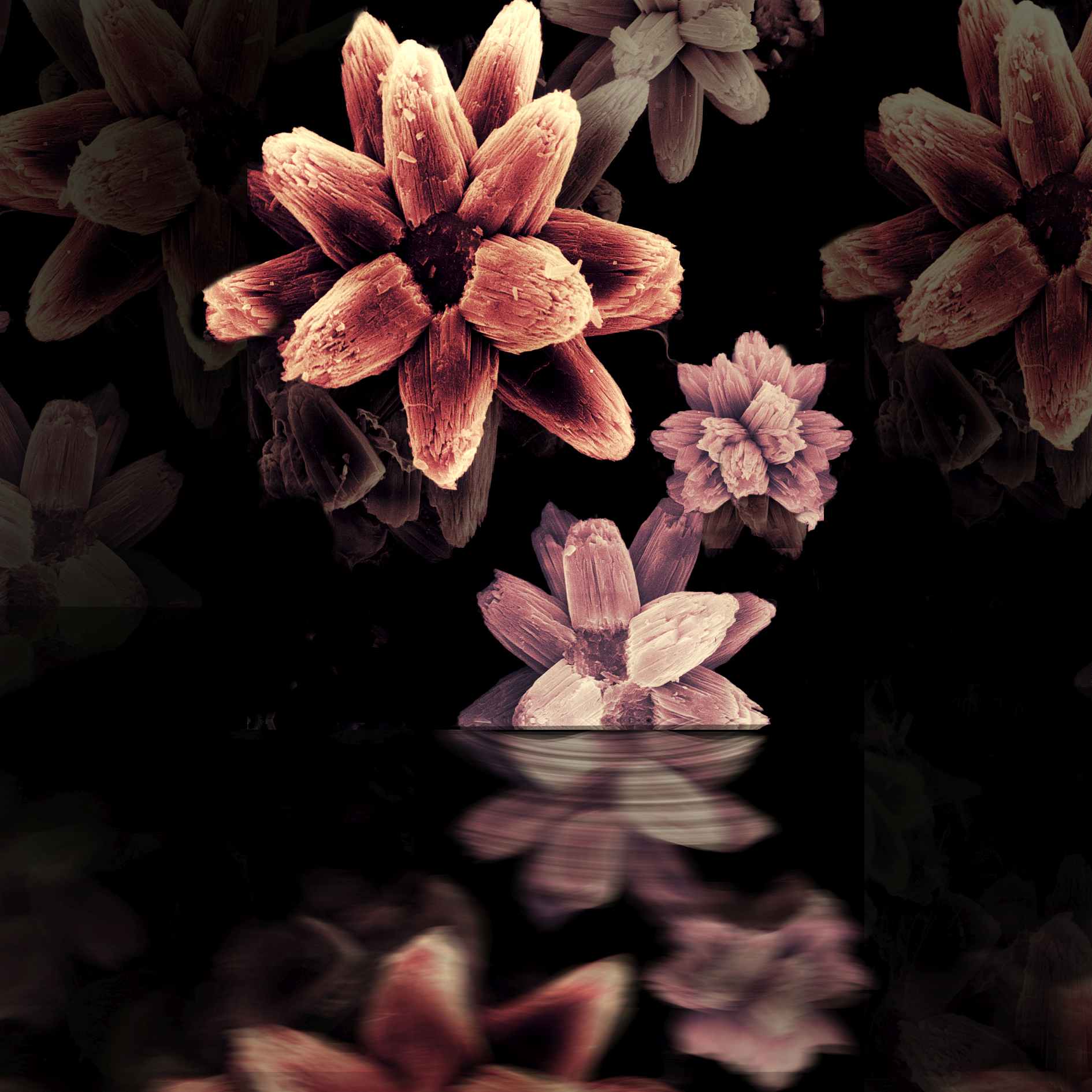
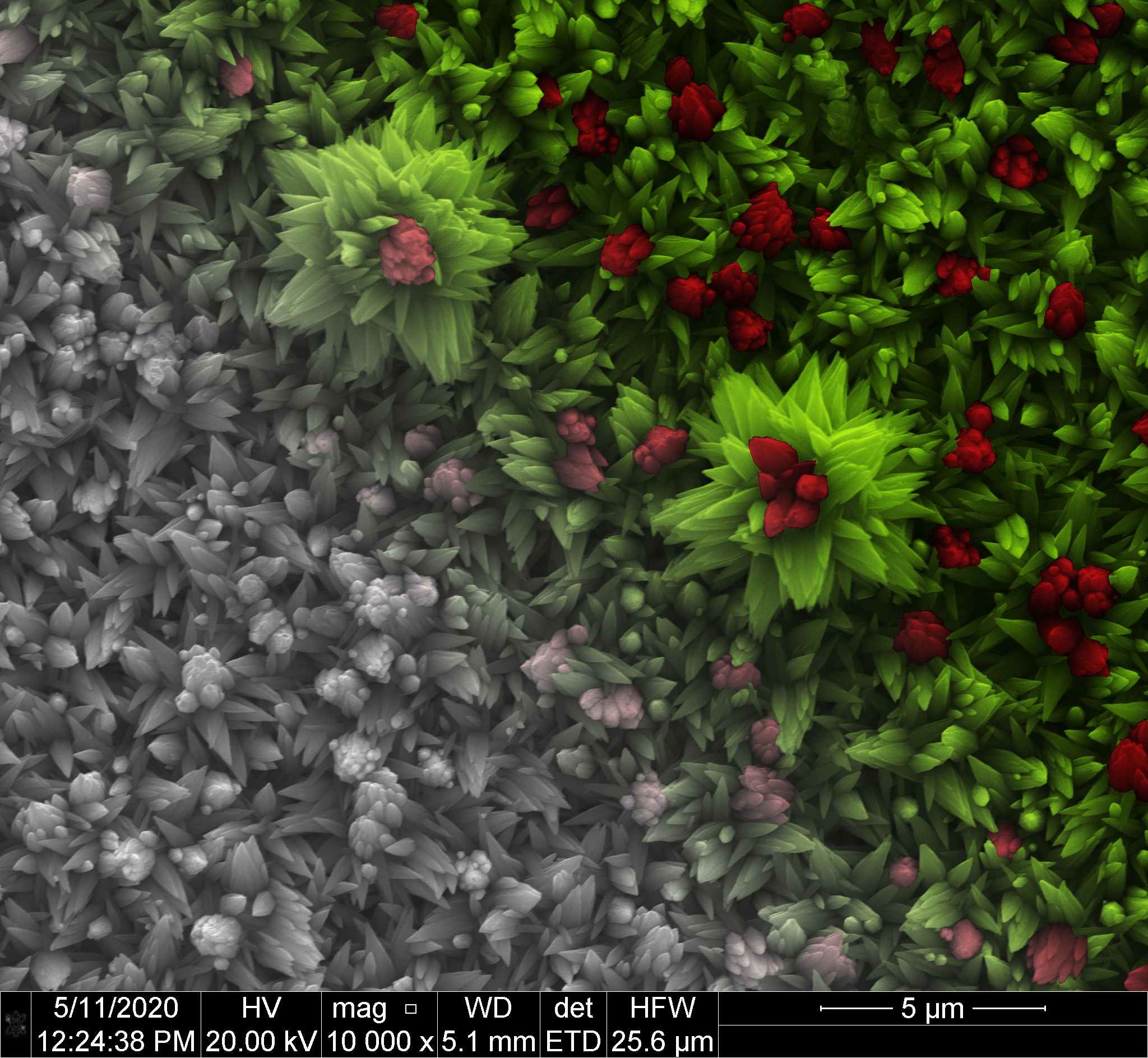 | Blossom of the Zinc Oxide Eitan Edri / Bar-Ilan University A SEM SE image showing a substrate covered by ZnO which grows in a flower-field form. |
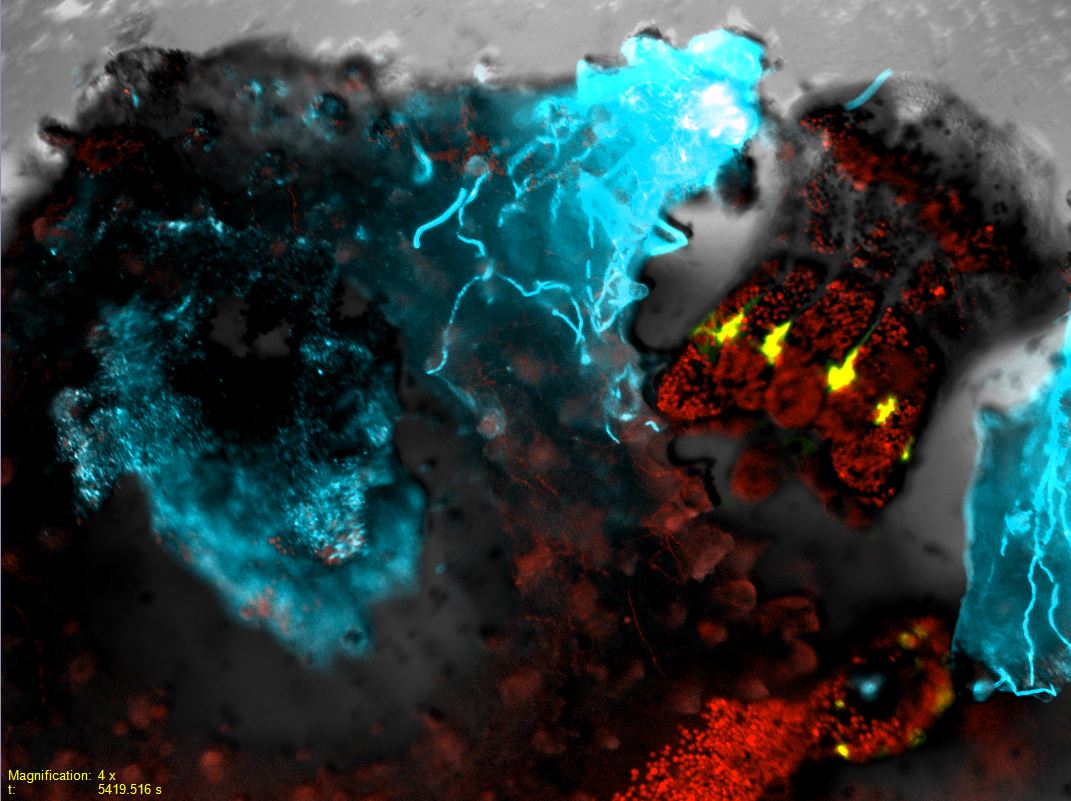 | Ascent to the Heavens Assaf Gavish / Weizmann Institute Light Microscopy image. As a colony of a reef-building coral (P. damicornis) is infected by its ruthless nemesis, the bacteria V.coralliilyticus, one courageous polyp decides to bail-out of his mothership, in hope of continuing her legacy in a safe haven.The bailed-out polyp, on the right, safe and sound with all its symbiotic algae (red – Chlorophyll) and tentacles presentingthe naturally occurring GFP (green), gazes upon the misfortune of his twin, a necrotic polyp (on the left), his bodyscavenged on by genetically-modified fluorescent bacteria (blue), revealing the skeleton of the colony on which it oncethrived (white). |
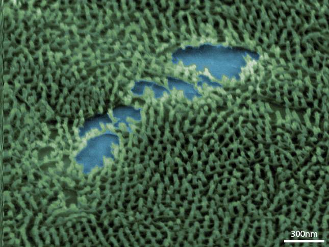 | Nano Pines Ofer Burg / Hebrew University A SEM image of an array of platinum pillars. The pillars resemble a dense forest or taiga. Digital colouring the empty areas in the middle of the image looks like lakes. |
 | Flatlands Rajashree Konar / Bar-Ilan University SEM SE mage of bulk Nickel Selenide (NiSe) grown in an ambient pressure CVD. |
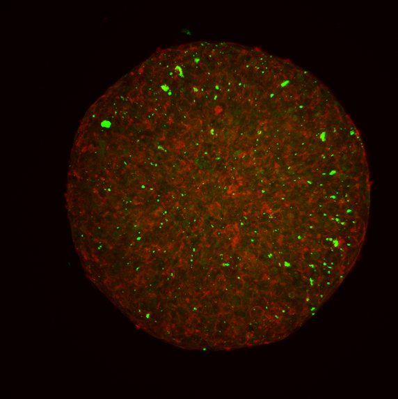 | Tumor Spheroids Treated With Particles Eliana Steinberg / Hebrew University A431 tumor spheroids stained with Actin and treated with particles loaded with Coumarin-6. Spheroids were cleared and then imaged with a Confocal Microscope. |
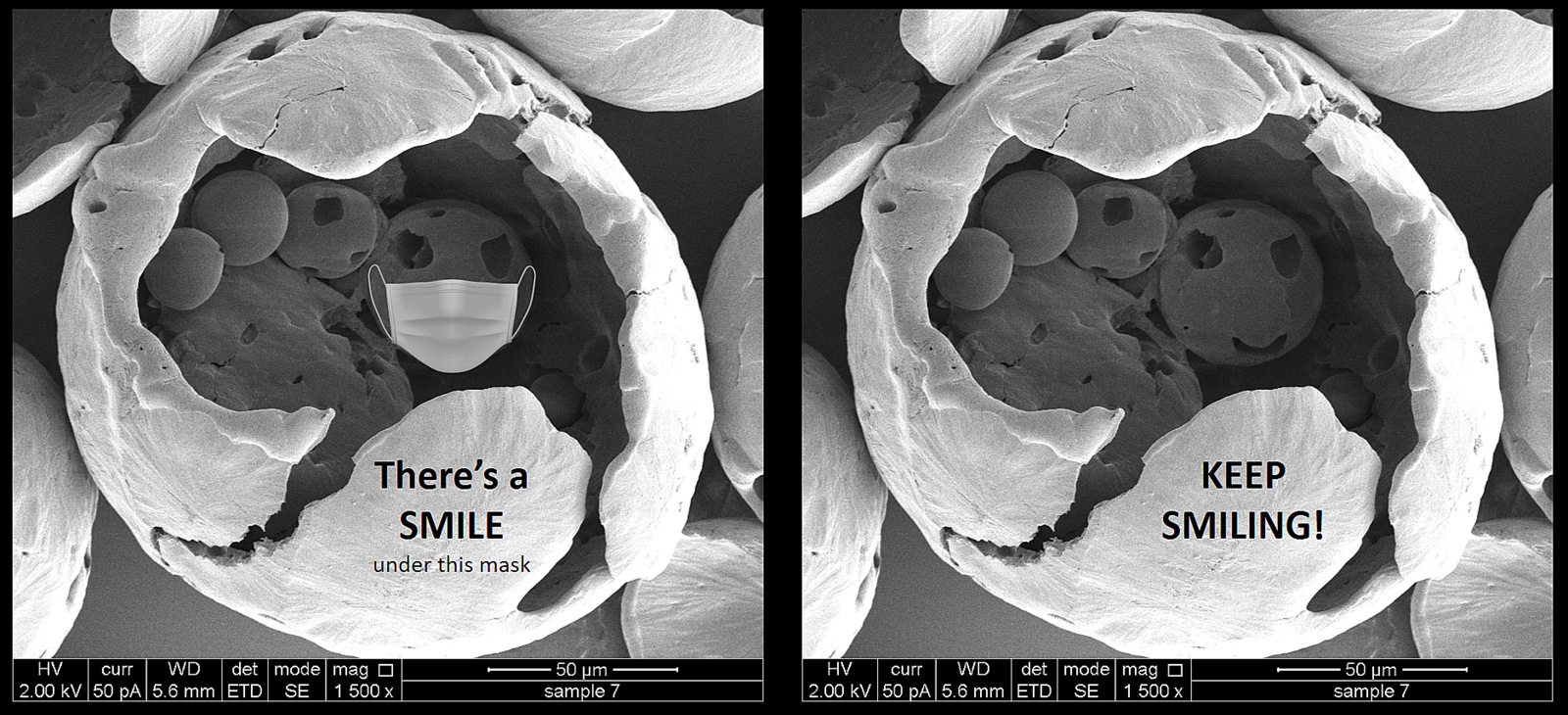 | Smile is the best medicine! Benzion Amoyav / Hebrew University The covid-19 pandemic has changed our world significantly, specifically the way we are socially interact. Keep smiling – with your mask on 🙂 The SEM SE image shows polycarpolactone (PCL) 12% particle prepared using a double emulsion method. |
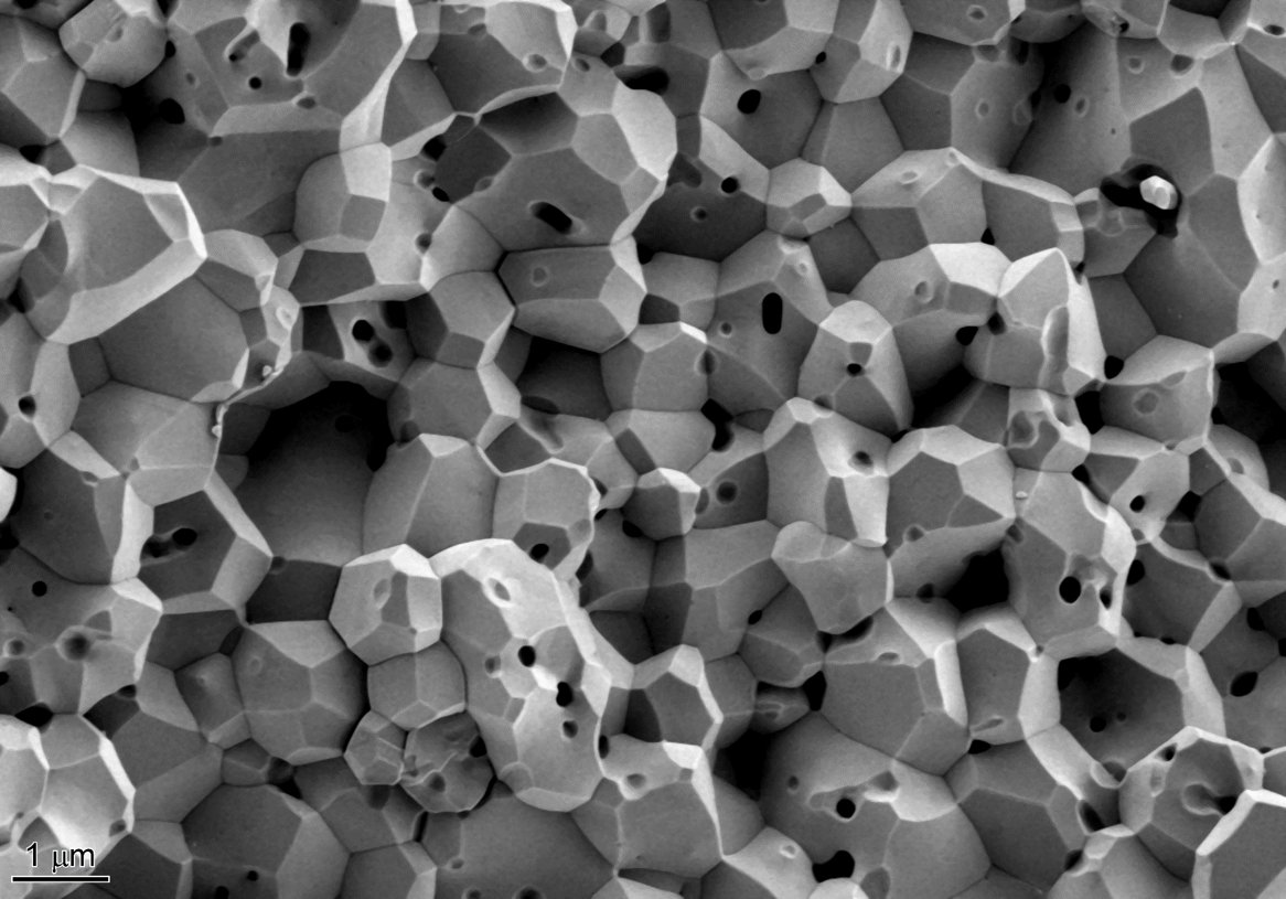 | Rapid Sintering Li-or Cohen / Technion The secondary electron micrograph was taken by Zeiss Ultra-Plus HRSEM microscope. The micrograph shows the microstructure of undoped alumina fabricated by slip-casting and rapid sintered at 1600⁰C for 3 minutes. The final body resulted in a relative density of 92%. |
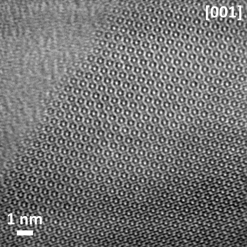 | CrPS4 Adam Budniak / Technion The edge of mechanically exfoliated CrPS4 in [001] Z.A. Please pay attention to the sharp, crystalline verge. X1M magnification micrograph registered by simple, thermionic gun, no-Cs-corrector FEI Tecnai T20 LaB6 TEM; ABSF (filtering) applied. |
 | Peptide self assembly Rotem Shitrit / Tel-Aviv university HydroxyProline-Phenylalanine-phenylalanine peptide self assembly on flexible surface - Optical microscopy. |
 | Nano Lotus ChanderPratap Singh / Ben-Gurion University SEM SE image of Vanadium sample. |
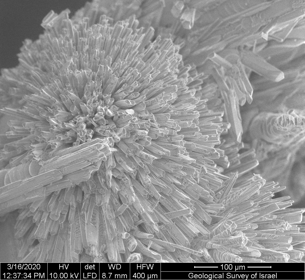 | Aragonite crystals Raanan Bodzin / Geological Survey of Israel SEM SE image of Aragonite crystals (one of the crystal forms of calcium carbonate CaCO3 mineral) from a hydrothermal cave in the northern Negev. Credit: Boaz Langford investigator, Raanan Bodzin - SEM. |
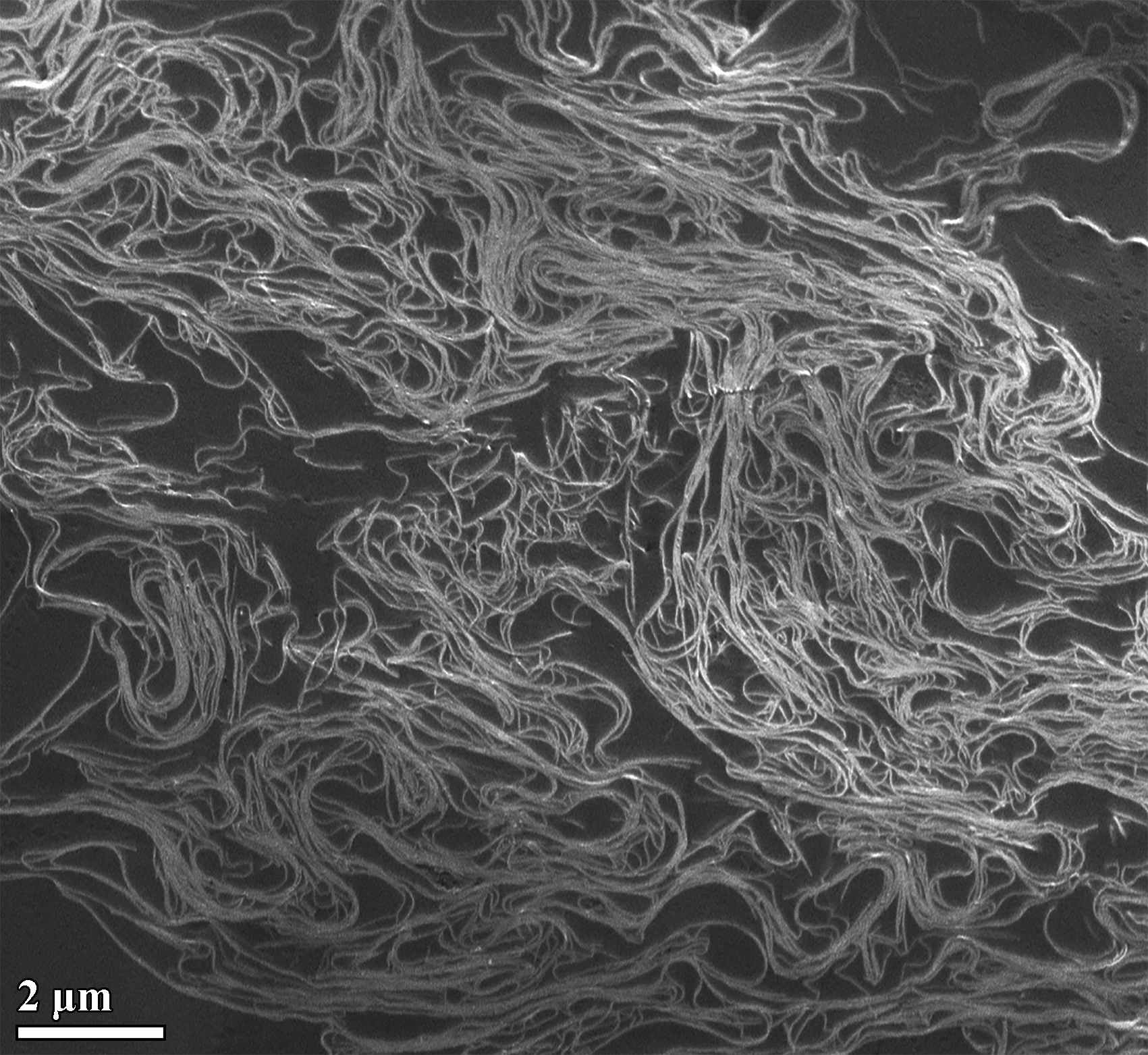 | Cryo-SEM of Carbon Nanotubes Asia Matatyaho-Yaakobi / Technion Cryo-SEM of carbon nanotubes (CNTs) dissolved in methanesulfonic acid at a concentration of 0.5 mg/mL showing an isotropic phase with short-range ordered domains. |
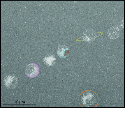 | Nano Space Linoy Dery / Hebrew University Self-assembly of silica nanoparticles on a surface looks like the solar system in SEM. |
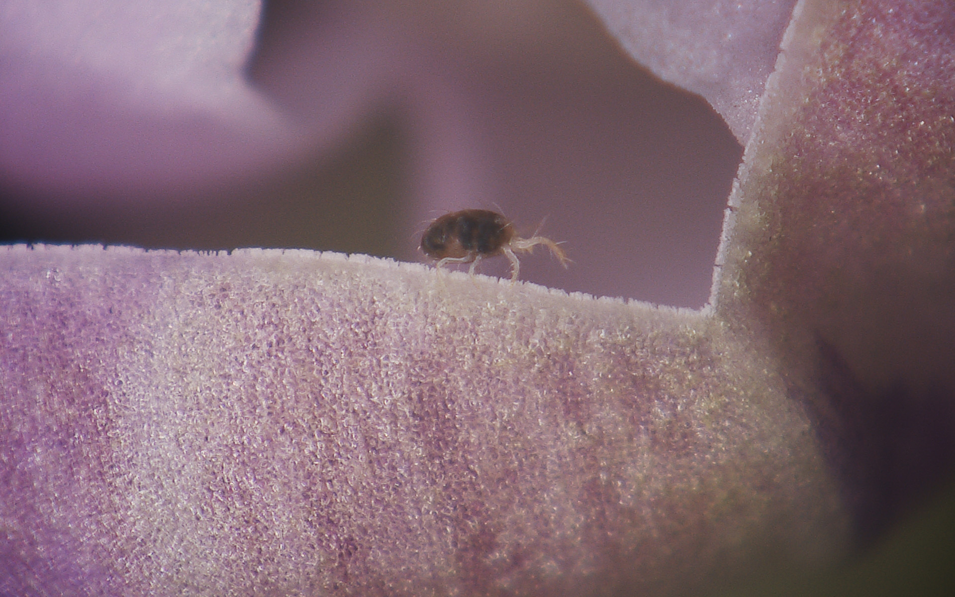 | Mite-Art: A Spider-Mite takes a hike Shira Gal / Volcani Center A Spider-Mite walking on a bean flower. Photo taken with a Hirox 3D digital microscope RH-2000. |