


| The 2025 Micrographs : |
 | 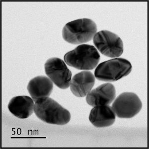 | MONUMENT (גל-עד) IN MEMORY OF ADI VITAL-KAPLOUN Z”L Adi Vital-Kaploun Ben Gurion University of the Negev Adi Vital-Kaploun Z”L, was an outstanding graduate of BGU's master’s program in desert studies, solar energy and environmental physics (Supervisor Prof. M. Y. Bashouti). Tragically she was brutally murdered on October 7th in Kibbutz Holit. This is an image of gold nanoparticles from her research, taken by Dr. Vladimir Ezersky on JEOL 2100F TEM. |
|
| 1 | 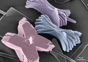 | BOWS & RIBBONS Zohar Eyal Weizmann Institute of Science Scanning electron microscopy image of synthetic guanine crystals. |
|
| 2 | 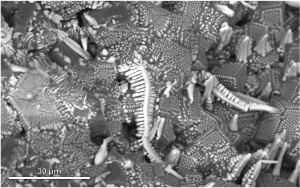 | METALLURGICAL CENTIPEDE Amram Azulay Tel Aviv University Scanning electron microscope micrograph of a fracture surface of a solidified bulk obtained by induction melting of Fe, V, and Al in a ceramic crucible. The image was acquired using backscattered electrons signal. |
|
| 3 | 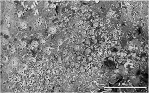 | FIELD OF WATER BEARS Hanna Bishara Tel Aviv University Scanning electron microscope micrograph of a fracture surface of a solidified bulk obtained by induction melting of Fe, V, and Al in a ceramic crucible. The image was acquired using a mix of backscattered and secondary electrons signal. |
|
| 4 | 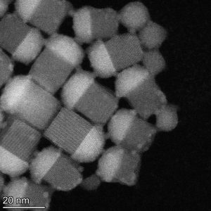 | NANO MUSHROMS Rachel Lifer Technion – Israle Institute of Technology HAADF STEM micrograph of epitaxial heterostructure quantum dots of Halide Perovskite and Lead Chalcohalide |
|
| 5 | 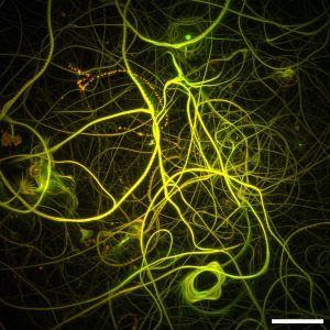 | MICROTUBULE HIGHWAYS Mohammed Aboraya Bar-Ilan university This image was taken using a Total Internal Reflection Fluorescence (TIRF) microscope and shows the movement of microtubules in a gliding assay. It’s a maximum intensity projection created by merging images captured over 30 minutes, allowing us to visualize the continuous motion and trajectories of the microtubules throughout the experiment. |
|
| 6 | 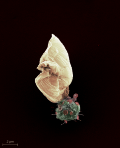 | OCEANIC BEAUTY Shay Kirzner Technion – Israle Institute of Technology This SEM image shows the marine unicellular microorganism Paraphysomonas bandaiensis. We can see its circular body covered in rounded scales that look like little umbrellas, colored in red. Attached to it, like a sale, is a sepia-colored structure that may be used as a shell. The grazer was collected with an Influx sorter, placed on poly-L-lysine coated silicon chips, critical point dried and coated with a layer of ~2nm of graphite. Samples then were viewed and captured using Zeiss Ultra Plus HR Scanning Electron Microscope. |
|
| 7 | 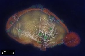 | UNDERWATER SENSU Shay Kirzner Technion – Israle Institute of Technology This SEM image shows the marine unicellular microorganism Paraphysomonas bandaiensis. We can see its circular body on the right, a fan-like structure and, colored in green, rounded scales that look like little umbrellas. The shape and size of the scales are used for identification of the species. Therefore, the word sensu in the title refers to the taxonomic term and also to the shape shown, of a traditional Japanese folding fan. The grazer was collected with an Influx sorter, placed on poly-L-lysine coated silicon chips, critical point dried and coated with a layer of ~2nm of graphite. Samples then were viewed and captured using Zeiss Ultra Plus HR Scanning Electron Microscope. |
|
| 8 | 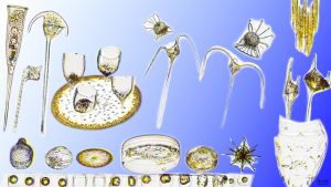 | MONARCH JEWELS & TOOLS Carmel Danino Gozlan University of Haifa Plankton images, captured using a PlanktoScope (a microscope with a flow cell and a camera). Among the organisms arranged in the picture are different species of dinoflagellates and diatoms. The organisms in the picture are in their original "royal" shapes and colors. |
|
| 9 | 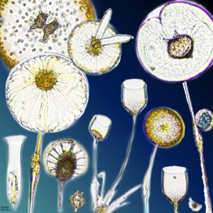 | PLANKTONIC GARDEN Carmel Danino Gozlan University of Haifa These live phytoplankton images were taken using a PlanktoScope (a microscope with a flow cell and a camera). Among the organisms arranged in the picture are different species of dinoflagellates and diatoms. The organisms in the picture are in their original colors |
|
| 10 | 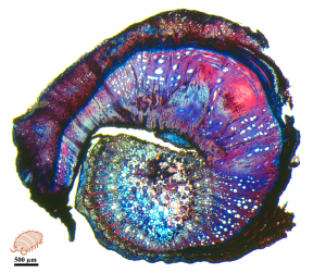 | A GRAPEVINE GRAFT UNION - OR A ROLY-POLY? Ilana Shtein Ariel University Light microscopy image of a grapevine graft union. Successful grafting unites two different plant species into one functional chimaera plant. Here we see an example of a failed union, without tissue fusion or new cambium formation, and with hormonal imbalance shown by circular abnormal growth of xylem cells. Histological sliding microtome section, Safranin-Alcian Blue stain. |
|
| 11 | 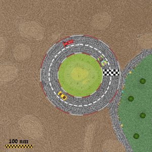 | FAST & VESICLES Sapir Rappoport Technion - Israel Institute of Technology The cryo-TEM micrograph shows multilamellar vesicles formed by amphiphilic interpolyelectrolyte complexes (IPECs) composed of poly(diallyldimethylammonium chloride) (PDADMAC) and poly(acrylic acid) sodium salt (NaPA), interacting with dioleoylphosphatidylcholine (DOPC) vesicles after three days. IPECs serve as promising drug delivery carriers, typically encapsulating drugs within their hydrophobic core. A key factor in IPEC-based drug delivery is the interaction between these carriers and lipid membranes, as it significantly impacts the efficient transport of therapeutic agents to intracellular targets. The image was taken using cryogenic transmission electron microscopy with Volta phase plate for image contrast enhancement. |
|
| 12 | 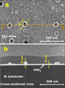 | HAFNIA GROWTH BY ATOMIC LAYER DEPOSITION TECHNIQUE TOP (A) AND CROSS-SECTIONAL (B) VIEWS Valentina Korchnoy Technion - Israel Institute of Technology Hafnia grows as a smooth polycrystalline small-grain structure, however sporadic grains with characteristic size of 60 nm are observed. Film thickness in smooth areas is ~20 nm. Hi-k dielectric deposition development for nanoelectronic applications. The micrograph was taken using ThermoFisher Helios 5 Dual Beam FIB-SEM instrument. |
|
| 13 | 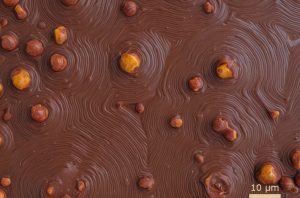 | SPIRALLING CHOCOLATE Larisa Popilevsky Technion - Israel Institute of Technology The contamination of the heat treatment oven led to the formation of the intricate structure. The micrograph was acquired using Dual Beam FIB-SEM (Helios G3, TFS). |
|
| 14 | 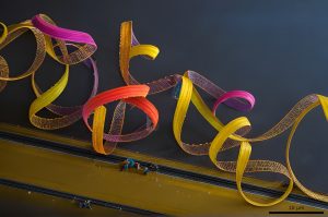 | MICRO-ROLLER COASTER Larisa Popilevsky Technion – Israel Institute of Technology The micrograph reveals the detachment of a gold (Au) layer from a cracked glass substrate. It was acquired using a Dual Beam FIB-HRSEM (Helios G3, TFS). |
|
| 15 | 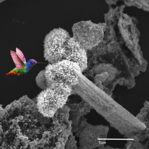 | MICROSCOPIC PARADISE Olga Krichevsky & Tamara Brider Ariel University Hight surface area of Catalyze materials including metal oxides and carbides. UH Res MAIA 3 FE-SEM, In-Beam SE detector. Mag 20kX.Scale bar 5micron. |
|
| 16 | 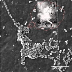 | "EVERY COLONY DREAM TO BE UNICORN" Olga Krichevsky & Tamara Brider Ariel University Ziv Dor, Prof. Shiri Navon-Venezia, E. coli on medical equipment. Bacterial Pathogenesis and Antibiotic Resistance Lab. UH-Res MAIA 3FE-SEM, Tescan. SE detector, Mag 10kX. Scale bar 10 micron. |
|
| 17 | 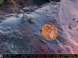 | FRACTURED YEAST Zipora Lansky & Olga Kleinerman Technion – Israel Institute of Technology CRYO-SEM image of Hybridized system of cellulose-coated oil in water emulsion with castor oil in the core with 1 wt.% yeast dispersion, Prof. Cohen and Ester Korkus. Taken on Zeiss UltraPlus HRSEM in the Technion Center for Electon Microscopy of Soft Matter |
|
| 18 | 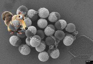 | GOLD AGE Chalom Zemmour Hebrew University of Jerusalem The image shows Janus hybrid nanoparticles (polymer-made particles partially covered in gold) which gives two properties to one same nanoparticle. The unique composition of this nanoparticle allows medicine inside it to be released in a specific area wherever the metal is being attracted to by a magnet, or in this case by Scrat, who thinks he finally got another acorn. |
|
| 19 | 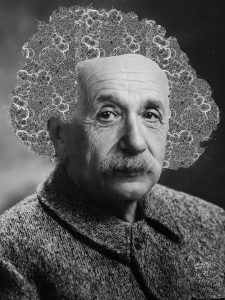 | BAD HAIR DAY Chalom Zemmour Hebrew University of Jerusalem Einstein is the muse in this representation of a wild lock of hair composed by a nanoscale net of porous titanium. This porous titanium nanomaterial’s shape enables a maximal absorption of drug substances. Just as the brain’s ability to seize knowledge in the ideal scientist. |
|
| 20 | 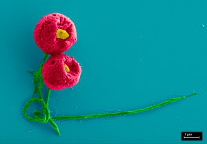 | ROMANCE UNDER THE LENS Maria Koifman Khristosov Technion – Israel Institute of Technology This unicellular nanoflagellate is called Paraphysomonas bandaiensis. It is known to graze on smaller phytoplanktonic cells, different bacteria and other particles in the oceans, and other aquatic environments. It has an important impact on the biogeochemical cycles of our planet. The sample was created by Shay Kirzner from Prof. Debbie Lindell group, Faculty of Biology, Technion. The micrograph was performed by Maria Koifman Khristosov using Zeiss Ultra-Plus FEG-HRSEM, Department of Materials Science and Engineering, Technion. Photoshop by Victor Khristosov. |


