


| The 2026 Micrographs : |
| 1 | 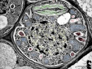 | Phloem transfer cell – a microcosmos of organelles Zohar Eyal Eastern R& D Center, Ariel University TEM image of a phloem transfer cell of Ifloga spicata, a small sand-trapping plant native to Israel. Phloem transfer cells are highly specialized parenchyma cells designed for intensive short-distance transport of sugars into or out of sieve elements of the phloem. They are distinguished by extensive cell wall ingrowths, which greatly increase membrane surface area to support high rates of transmembrane transport. |
|
| 2 | 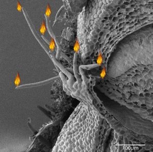 | CANNUKAH MENORAH IN A MANGO (MANGIFERA INDICA) LATERAL BUD Tamara Brider, Avishai Londner, Ilana Shtein Ariel University The image was taken with a TESCAN Maia3 microscope, using an SE detector. Before testing, the sample was prepared according to a standard method and dried by CPD. |
|
| 3 | 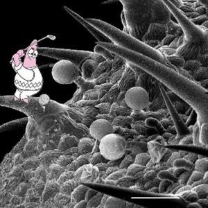 | GOLF ON SAGE LEAF Olga Krichevsky Ariel University Glandular and non-glandular trichomes on a sage (Salvia) leaf. Sage (Salvia) leaf exhibits several types of glandular trichomes that act as "bio-factories" for essential oils and metabolites and offer chemical defense against herbivores. In addition, non-glandular defensive trichomes offer mechanical protection to the leaf. The sample was dried in CPD and gold coated. UHR MAIA 3 FE-SEM (Tescan), SE detector, Scale bar 100 micrometer. Avishai Londner , Ilana Shtein lab |
|
| 4 | 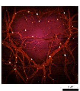 | SILKEN THORNS Mariia Rodionova Weizmann Institute of Science This image shows a silk nanofibrillar network. The sample was prepared from a regenerated silk solution for characterization of an experimental attempt to mimic a nanofibrillar solution of native silk fibroin, found in the final part of the gland of Bombyx mori silkworm. The image was obtained using Atomic Force Microscopy with a JPK Nanowizard 4 and processed in Gwyddion software. The sample was drop-cast onto a silica wafer, which was previously activated with oxygen plasma to improve hydrophilicity. |
|
| 5 | 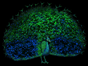 | FROM KERTINOCYTES TO FEATHERS Susanna Feldman Oncology, Shamir Medical Center This image merges immunofluorescence microscopy with a familiar display from the animal world. Human skin keratinocytes were stained for the immune checkpoint ligand OX40L (green) and counterstained with DAPI (blue), revealing a cellular architecture that immediately reminded us of a peacock’s tail fan. Coincidentally, a real male peacock has been wandering around our hospital grounds for several years, delighting staff and patients and earning the name “Yossi.” Inspired by this local celebrity, we photographed Yossi displaying his magnificent tail and replaced the feathers with our microscopic “peacock.” The result connects similar patterns across biological scales, turning immune signaling into feathers and reminding us that scientific discovery often comes with unexpected moments of beauty—and a touch of humor—from the natural world. |
|
| 6 | 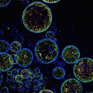 | SPACE-TIME FOAM Alexandra Tsitrin & Yael Cohen Ilse Kats Institute of Nanoscale Science and Technology, Ben Gurion University of the Negev Super resolution microscopy (SIM2), x20 magnification Object: synthetic peptide polymerization and formation of micro bubbles |
|
| 7 | 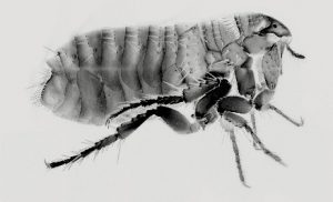 | HOOKE’S FLEA, 360 YEARS LATER (HOMAGE TO ROBERT HOOKE) Alexandra Tsitrin Ilse Kats Institute of Nanoscale Science and Technology, Ben Gurion University of the Negev Confocal microscopy of chitin autofluorescence. Ctenocephalides felis, the common cat flea, was first depicted in detail by Robert Hooke in his groundbreaking book Micrographia (1665). This illustration is considered one of the earliest truly scientific images of a microscopic animal in human history. It set new standards for accuracy, observation, and visual communication, helping to establish scientific illustration as a powerful tool for both research and the popularization of science. More than 360 years later, modern imaging allows us to revisit the same tiny organism and discover its intricate beauty from entirely new perspectives—bridging centuries of scientific curiosity and technological progress. |
|
| 8 | 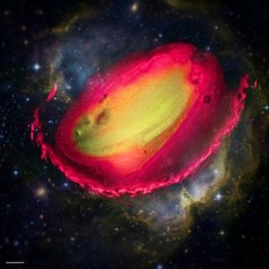 | COSMIC SEED Etty Grad Bar Ilan University Delivery of Exogenous GFP (DNA) to Arabidopsis thaliana wild type seed via Hydrogel nano-particles. Vladislav Rapaport, Gad Miller's lab. Please include in the legend that the scale bar represents 50 µm and that the image was taken using a 20× objective. |
|
| 9 | 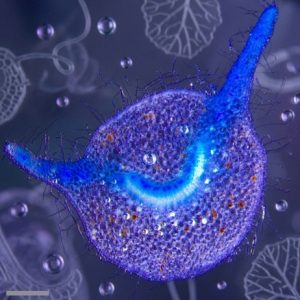 | DON'T WORRY BE HAPPY! Irit Shoval Bar Ilan University Fom-1 promoter:GUS constructs was introduced into Agrobacteriumtumefaciens and stable transgenic Nicotiana tabacum plants were generated. Tissue cross sections of leaf petiole were stained for GUS activity and observed under the Leica M205 Streoscope. Dr. Amalia Bar-Ziv from Prof. Rafael Perl-Treves lab scale bar=500um. |
|
| 10 | 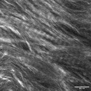 | A BONE TO PICK Avital Wagner Radboud University Medical Center Bright-field scanning transmission electron microscopy (STEM) image of a thin bone lamella prepared using a focused ion beam/scanning electron microscope (FIB/SEM). In the image the two main components of bone are visible: a scaffold of collagen fibrils with their characteristic 67 nm banding pattern and intrafibrillar 4 nm-wide calcium phosphate mineral platelets. |
|
| 11 | 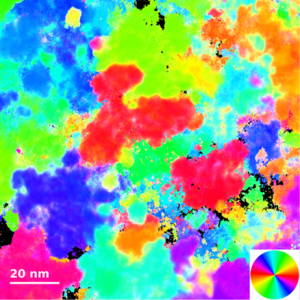 | NANOCRYSTALLINE CLOUDS AT THE END OF THE RAINBOW Anastasiya Reveguk Tel Aviv University A fragment of dark-field TEM orientation map of a nanocrystalline Fe3Si thin film. The coloring is based on angular positions of the {110} and {211} reflections (color wheel in the insert). |
|
| 12 |  | PLANKTON PARTY Irit Shoval Bar Ilan University Diatoms produce about 1/5 of the oxygen we breath. Different growing conditions (resource limitation, viral predation) of Chaetoceros tenuissimus, a single-cell diatom species, results in different morphologies. Autofluoresecnce chlorophil (red) and Diatome silica wall was stained with PDMPO (green). Images were acquired on Leica Stellaris 5 confocal microscope using the 63x oil objective. Scale bar = 10um. Ori Zucker and Yael Tennenbaum-Azulai, Kranzler lab. |
|
| 13 | 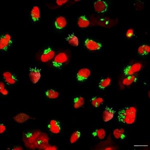 | STRAWBERRY FIELDS FOREVER Irit Shoval Bar Ilan University This image shows kidney cells expressing different mutations in the renin protein, a key regulator of blood pressure and kidney physiology. To highlight cellular architecture, the Golgi apparatus was labelled with GFP, while the nucleus was labelled with mScarlet. The cells were imaged live using a confocal spinning disk microscope (Opera Phenix Plus High Content Screening System) with a 40× water-immersion objective. The fluorescent labelling reveals clusters of Golgi structures surrounding brightly marked nuclei, creating a pattern reminiscent of a field of strawberries. Scale bar: 25 µm. Narkis Arbeli, Moran Dvela Levitt Lab. |
|
| 14 | 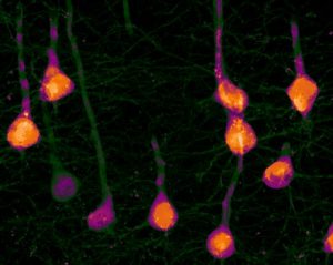 | NEURONAL POP ART Pratibha Ahirwal Technion – Israel Institute of Technology Corticospinal neurons in the mouse motor cortex labeled via retro-AAV tdTomato injection into the spinal cord (C6–C8). The image was acquired using spinning-disk confocal microscopy with a 60× objective, 560 nm laser excitation at 20% power, and 200 ms exposure. It is presented as a maximum intensity projection with uniform contrast adjustment. |
|
| 15 | 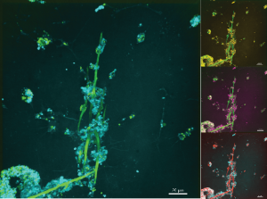 | A LIVING SCAFFOLD VESICLES WEAVING THE SKELETON Prashant Tewari University of Haifa A bright, sea urchin spicule runs through the center, while small glowing vesicles in skeletogenic cells move around it. These vesicles are moving in the cells along the structure and especially near the tip, where growth is happening. The image shows how skeletogenic cells come together to build a larger, stable structure. Sample: Sea urchin skeletogenic cell culture with spicule. Microscopy: Spinning Disc Confocal Microscope. |
|
| 16 | 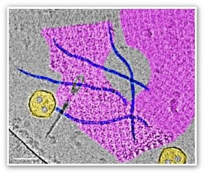 | NANO-FABRIC OF LIFE Ellina Kesselman Technion – Israel Institute of Technology This cryo-TEM image shows bacteriophages released from bacterial isolates after treatment with mitomycin C, which was used to trigger prophages inside the bacteria. The scale bar is 50 nm. The sample was filtered, concentrated, and vitrificated to preserve the natural structure of the viruses. The image was taken by Dr. Ellina Kesselman, Technion Center for Electron Microscopy of Soft Matter, The Wolfson Department of Chemical Engineering, Technion. The research was performed by Vibhaw Shrivastava, PhD Candidate, guided by Prof. Naama Lang-Yona, in the Atmospheric & Environmental Microbiology Lab of the Civil & Environmental Engineering Faculty, Technion. |
|
| 17 | 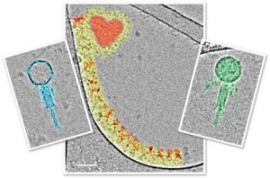 | NANO-WORLD PARTY Ellina Kesselman Technion – Israel Institute of Technology These cryo-TEM images show bacteriophages released from bacterial isolates after treatment with mitomycin C, which was used to trigger prophages inside the bacteria. The scale bar is 50 nm. The samples were filtered, concentrated, and vitrificated to preserve the natural structure of the viruses. The images were taken by Dr. Ellina Kesselman, The Technion Center for Electron Microscopy of Soft Matter, The Wolfson Department of Chemical Engineering, Technion. The research was performed by Vibhaw Shrivastava, PhD Candidate, guided by Prof. Naama Lang-Yona in the Atmospheric & Environmental Microbiology Lab of the Civil & Environmental Engineering Faculty, Technion. |
|
| 18 | 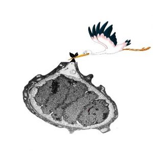 | A SPEEDY DELIVERY AT 11,000x Shivani Pundir Technion – Israel Institute of Technology Five cap cells unwrapped within a shared ECM from Drosophila Melanogater's lateral pentascolopidial organ. |


